I think everyone can relate to packing problems – how to get a cart full of groceries into the fewest bags or how to strategically organize the needed clothes and accessories into one carry-on suitcase? Packing problems abound in life from our personal needs to manufacturing concerns about how to fit the most items into the shipping box. There is a whole science to packaging with equations, theories, and even college degrees. Yet most of these macro-scale problems are almost trivial compared to the packaging problem inherent in every one of our cells. The average human cell is between 20-30 micrometers in diameter, roughly one-thousandths of an inch, a size only visible with a microscope. As small as cells are, within each of our cells (except red blood cells and certain cornified cells of the skin, nails, and hair) is our entire DNA genome. Although DNA is a very skinny molecule that is only 1 ten-millionth of an inch in diameter, it is an incredibly long molecule. If you take all the DNA in your 23 pairs of chromosomes from one cell and stretch it out end-to-end it would be roughly six and a half feet long, and all of that must fit inside a microscopic cell. And in fact, the DNA actually resides within the nucleus of the cell which is about half the size of the entire cell. This means that DNA must be compacted to roughly 150,000 times its original length. Getting this huge length of DNA into this microscopic container is a packaging nightmare that far exceeds our modest real-world packaging problems.
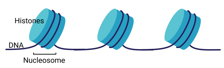
While science is a long way from a complete understanding of DNA packaging, many important players in the packaging apparatus have been identified. The first level of packing is the association of DNA with proteins called histones. The histones assemble into cylindrical structures and DNA wraps around the outside of these histone bodies to form nucleosomes (Fig. 1).
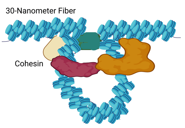
This “beads-on-a-string” stage of DNA is further compacted by coiling the nucleosome chain into the so-called 30-nanometer fiber (imagine wrapping beads on a string tightly around a pencil) (Fig. 2).

These 30-nanometer fibers are then folded into loops that are more and more compacted until the entire 6.5 feet of DNA can fit within the tiny cell nucleus. This remarkable packing is orchestrated by additional protein complexes, including one called cohesin. The cohesin complex consists of four different proteins that form a ring-like structure (Fig. 3).
Because of this ring structure, it was believed that cohesin wrapped around the 30-nanometer fiber to extrude loops. However, a recent publication in the journal Cell challenges that model. Using sophisticated molecular imaging techniques and various cohesin mutants, the researchers showed that the cohesin complex has two functional sites, the hinge and the head. First, the hinge binds the DNA and then the protein bends to pass the DNA to the head. Once the DNA is anchored to the head the hinge releases, swings back, and grabs another stretch of DNA. This newly bound DNA is passed to the head as the original head-bound DNA is extruded into the growing loop. The combined process resembles the action of pulling a rope hand-over-hand where one hand is the hinge and the other hand is the head. While this new model of cohesin action may be of interest mostly to the scientific community rather than the general public, this work is critical for understanding the intricate workings of our genomes as our DNA folds and unfolds continuously during the life of each cell. Genetic defects in DNA packaging are associated with several significant diseases such as alpha thalassemia X-linked intellectual disability syndrome, Coffin-Lowry syndrome, Rett Syndrome, Rubinstein-Taybi syndrome, and certain cancers. The greater our knowledge of this complex process the greater our ability to someday address the disease consequences of aberrant DNA packaging.

Leave a comment